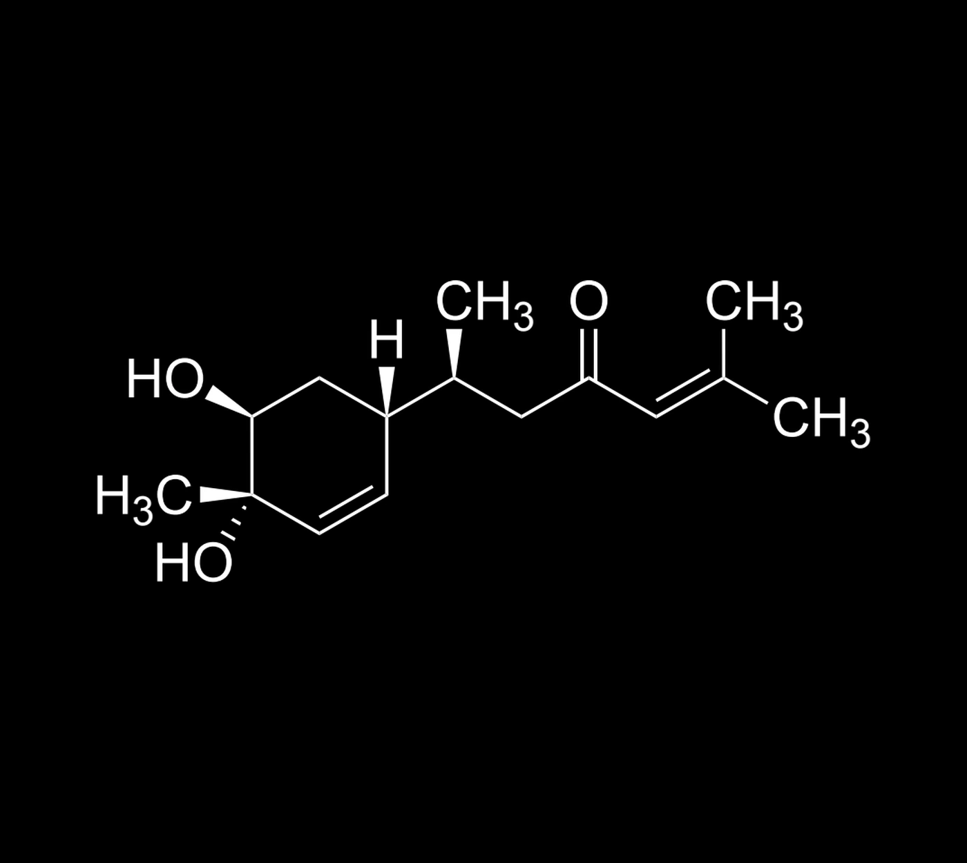
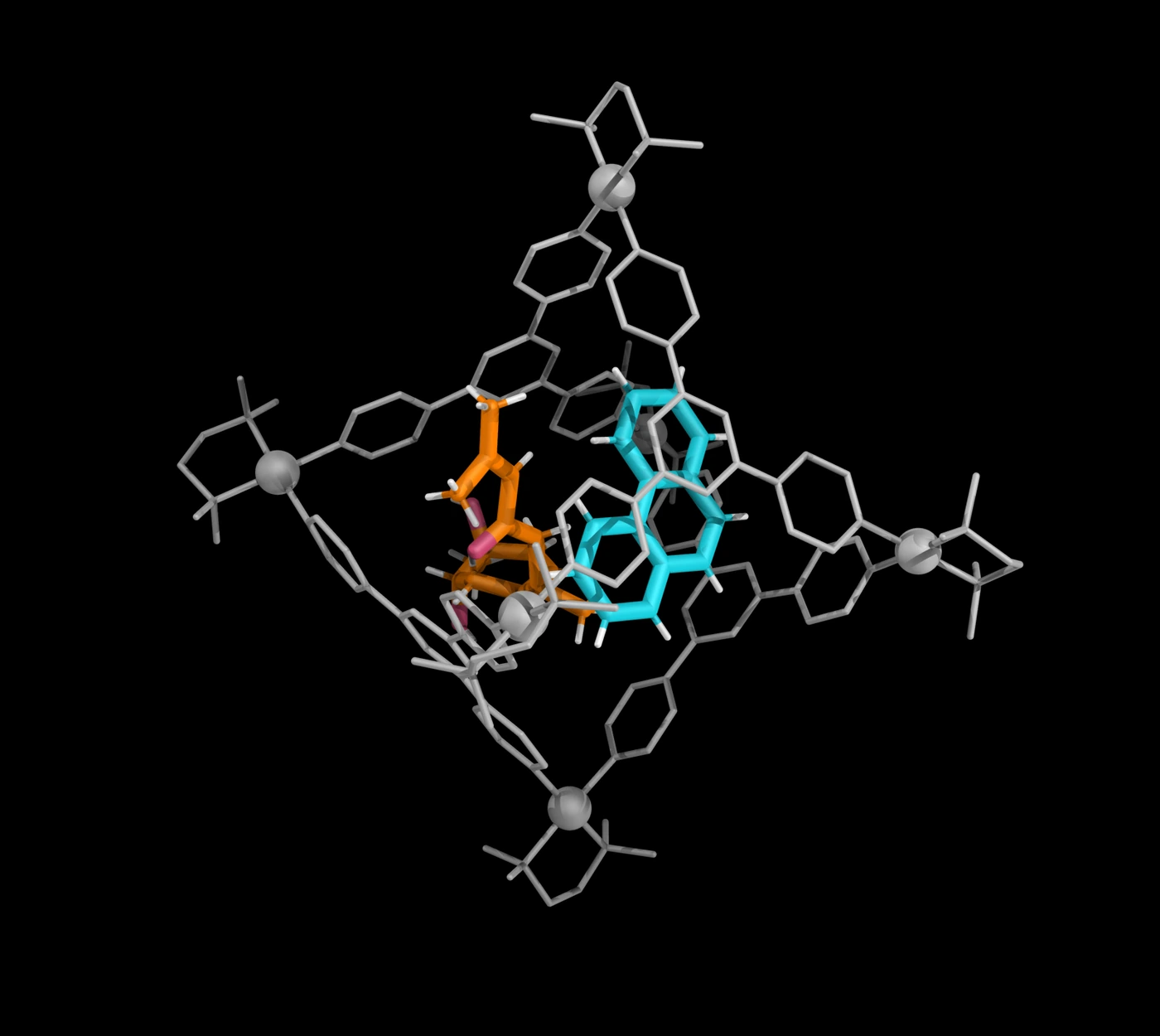
Bisacurone (compound 25) was structurally analyzed using cage 1a and phenanthrene (2a) in the size-compensation strategy for low-polarity small molecules.
G095 · Sesquiterpenoid
Bisacurone
C15H24O3 · 252.35 g mol−1
First crystal structure · Reported for the unmodified analyte in this study → · Read the story ↓
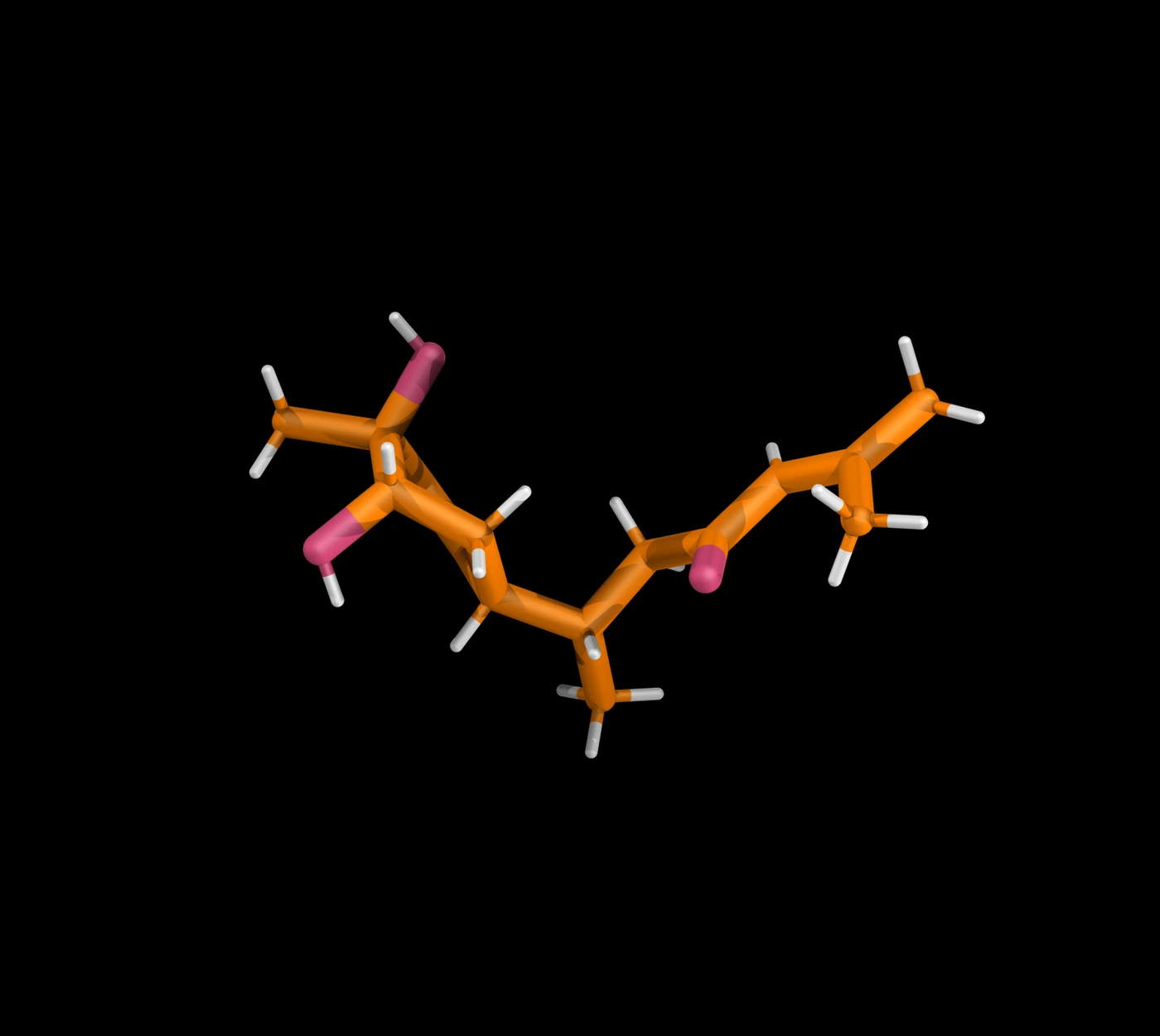
INTERACTIVE 3D STRUCTURE
Explore Bisacurone in its crystalline sponge
Drag to rotate, scroll to zoom, switch the cage on or off, and export the current view.
CHONCagePhenanthrene 2a
- Molecular formula
- C15H24O3
- Molecular weight
- 252.35 g mol−1
- CCDC accession
- 2565750 ↗
- Host
- Pd6L4 cage 1a
- Compound in publication
- 25
MOLECULAR HISTORY
From a newly isolated bisabolane to a crystal structure
Bisacurone was introduced as a new constituent of Curcuma xanthorrhiza, with its structure assembled from spectroscopy, chemical conversions and chiroptical evidence. Later cell studies gave researchers additional reasons to investigate the molecule. The present work adds a crystal model of the unchanged compound to this earlier chemical and biological record.
- 1989
Isolation and an absolute structural assignment
Uehara and colleagues isolated bisacurone with two other new bisabolane sesquiterpenoids from Curcuma xanthorrhiza rhizomes. Spectroscopy, chemical conversions and the exciton chirality method were combined to assign their absolute structures. Bisacurone therefore entered the literature with a stereochemical model, rather than as an unidentified extract component.1
- 2008
The isolated molecule is studied in cells
Sun and colleagues examined bisacurone in cultured human endothelial cells stimulated with TNF-α. They observed reduced VCAM-1 expression and less adhesion by monocytes and the cancer cell lines they tested. The isolated sesquiterpenoid had become a subject of cell-based studies of inflammatory adhesion.2
- 2026
A host–guest crystal supplies a molecular view
The published size-compensation study places bisacurone and phenanthrene together inside cage 1a. X-ray analysis resolves the chemically unmodified guest in the resulting crystal, which the authors report as its first crystal-structure characterization. This adds structural observation without replacing the original spectroscopy and chiroptical assignment.3
WHY THIS STRUCTURE MATTERS
Bisacurone entered the literature through spectroscopy, chemical transformations and chiroptical evidence. Its first reported crystal structure now shows the chemically unmodified molecule within the host, adding a three-dimensional diffraction model to that earlier structural assignment.
References
- Uehara, S.; Yasuda, I.; Takeya, K.; Itokawa, H. “New bisabolane sesquiterpenoids from the rhizomes of Curcuma xanthorrhiza (Zingiberaceae).” Chemical and Pharmaceutical Bulletin 1989, 37, 237–240. DOI 10.1248/cpb.37.237 ↗
- Sun, D.-I.; Nizamutdinova, I. T.; Kim, Y. M.; et al. “Bisacurone inhibits adhesion of inflammatory monocytes or cancer cells to endothelial cells through down-regulation of VCAM-1 expression.” International Immunopharmacology 2008, 8, 1272–1281. DOI 10.1016/j.intimp.2008.05.006 ↗
- W. He; L. Soliman; H. Takezawa; M. Fujita. “Expanding the Chemical Space of Analytes in the 2G Crystalline Sponge Method: Size-Compensation Strategy for the Structural Analysis of Low-Polarity Small Molecules.” Angew. Chem. Int. Ed. 2026, e4748505. DOI 10.1002/anie.4748505 ↗
Crystal records in this study
| Host | Modulator | CCDC | SI table |
|---|---|---|---|
| Standard cage 1a | Phenanthrene 2a | 2565750 | S22 |
SOURCE PUBLICATION
Expanding the Chemical Space of Analytes in the 2G Crystalline Sponge Method: Size-Compensation Strategy for the Structural Analysis of Low-Polarity Small Molecules
W. He, L. Soliman, H. Takezawa, M. Fujita
Angew. Chem. Int. Ed. 2026, e4748505.
First published: 20 October 2026 · Early View
DOI 10.1002/anie.4748505 ↗